Direction 1: Visualize and study single molecular tensions in cells
Utilizing CFN (cellular force nanoscopy) developed in our lab, we can literally see single molecular force (the bright sparks in the video) in live cells. Based on the imaged molecular tensions, we achieve super-resolution cellular force imaging by molecule localization. CFN provides an cutting-edge imaging tool for the study of force-structure interplay in live cells with ultra sensitivity and resolution. We are applying CFN to study the force and structural formation in some smallest adhesion units in cells.
Direction 2: Study cell mechanobiology of platelets
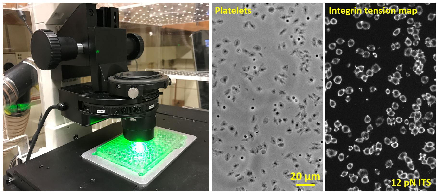
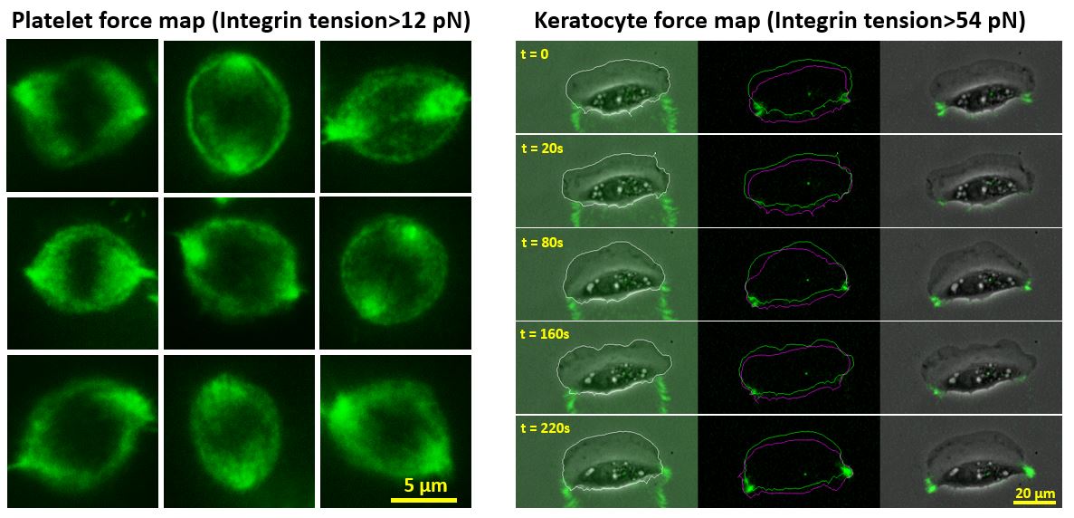
By converting force signal to fluorescent signal, integrative tension sensor (ITS) enables cellular force mapping directly by fluorescence imaging, therefore inheriting many advantages of fluorescence microscopy such as high resolution, high sensitivity and rapid image acquisition. With ITS, we are able to calibrate and map integrin tensions in real time in live cells such as platelets and migrating keratocytes. The cellular force mapping with high temporal and spatial resolution helps us explore the roles of integrin tensions in platelet adhesion, contraction and keratocyte protrusion and retraction. We are also studying the relation between platelet force maps and platelet health conditions, in the hope of developing a platelet force assay to assess bleeding risk in patients.
Direction 3: Harness cell mechanotransduction by molecular tension control
It is well recognized that cells are able to sense mechanical properties of the matrix and regulate their functions accordingly. Tension gauge tether (TGT) enables quantitative control of cellular force at the molecular level. TGT globally restricts molecular tensions on mechano-sensitive receptors such as integrins under a designed level Ttol, providing a unique tension knock-down technique.