|
Representative
Publications
|
|
Monitoring Escherichia coli in Water through Real-Time Loop-Mediated Isothermal Amplification on Biochips
|
(Micromachines 2024)
|
|
Photothermal Utility Heating with Diffused Indoor Light via Multiple Transparent Fe3O4@Cu2-xS Thin Films
|
(Energy Technology 2024)
|
|
A photothermal solar tunnel via multiple transparent Fe3O4@Cu2-xS thin films for heating utility application
|
(Solar Technology 2024)
|
|
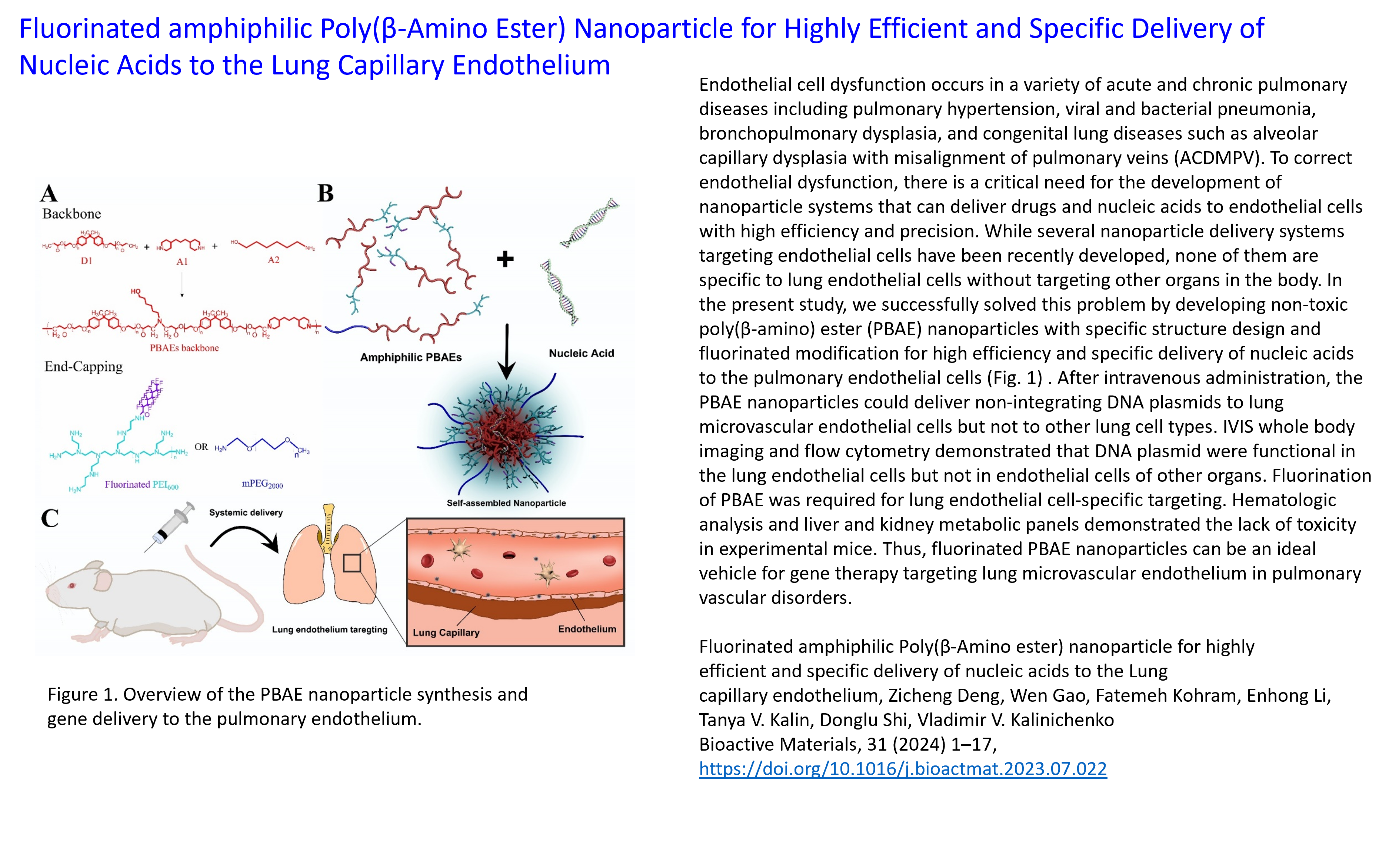
Fluorinated amphiphilic Poly(??-Amino Ester) Nanoparticle for Highly Efficient and Specific Delivery of Nucleic Acids to the Lung Capillary Endothelium
|
(Bioactive materials 2024)
|
|
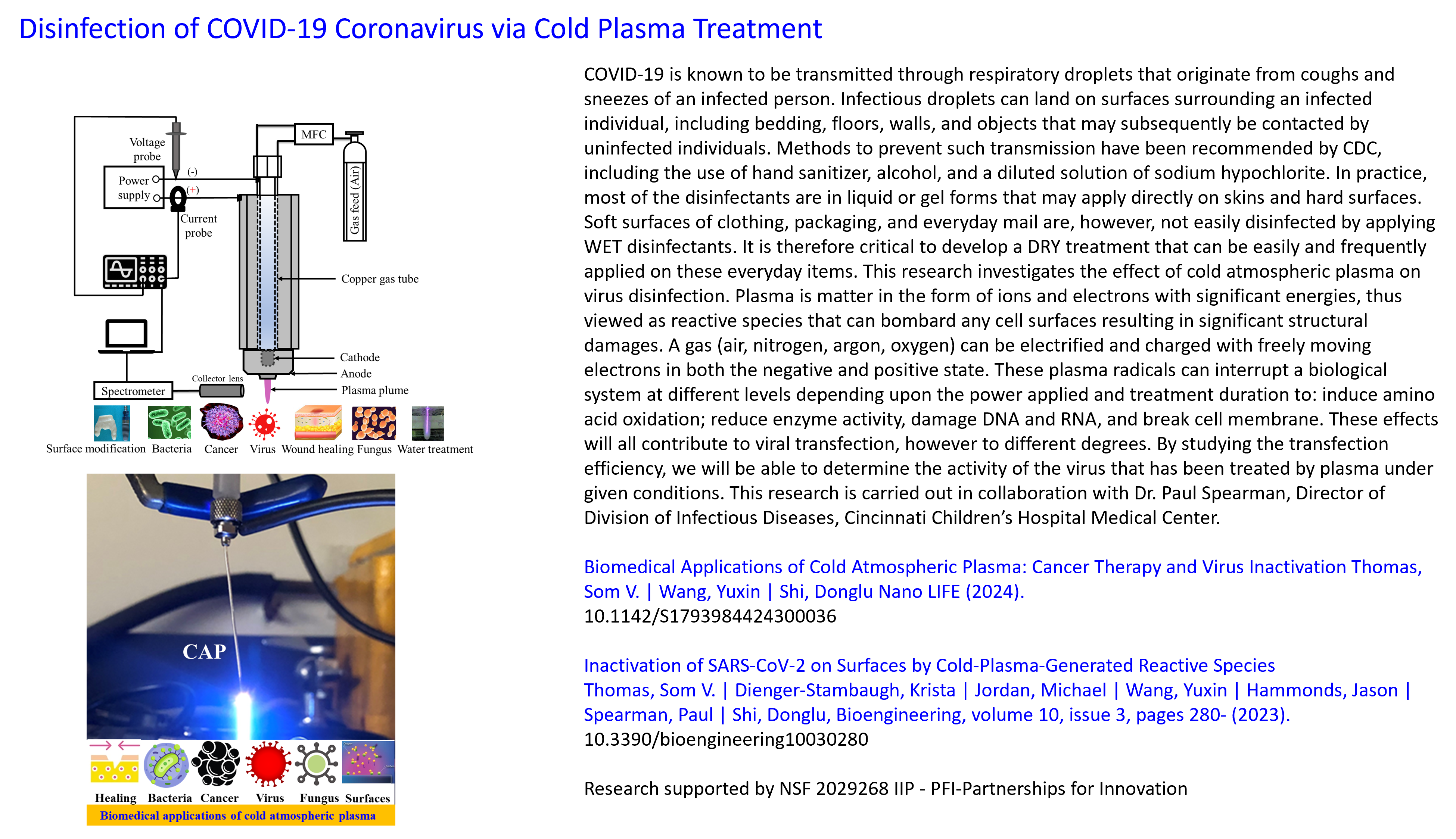
Biomedical Applications of Cold Atmospheric Plasma: Cancer Therapy and Virus Inactivation
|
(Nano LIFE 2024)
|
|
Solar harvesting through multiple semi-transparent cadmium telluride solar panels for collective energy generation
|
(Solar Energy 2023)
|
|
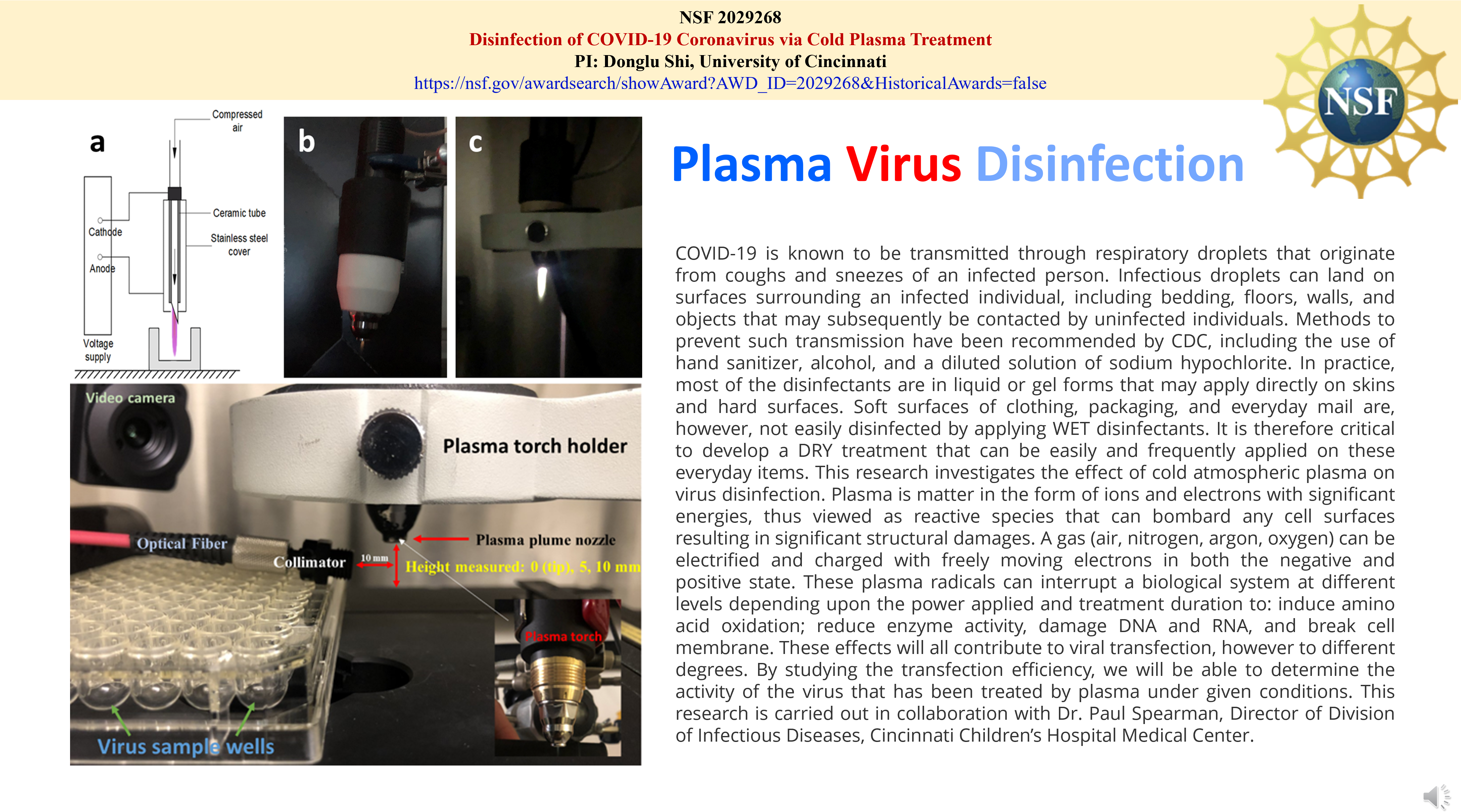
Inactivation of SARS-CoV-2 on Surfaces by Cold-Plasma-Generated Reactive Species
|
(Bioengineering 2023)
|
|
Lung endothelial cells regulate pulmonary fibrosis through FOXF1/R-Ras signaling
|
(Nature Communications 2023)
|
Demonstration of Safety in Wild Type Mice of npFOXF1, a Novel Nanoparticle-Based Gene Therapy for Alveolar Capillary Dysplasia with Misaligned Pulmonary Veins
|
(Biologics: Targets and Therapy 2023)
|
Enhanced Silicon Photovoltaic Efficiency by Solar Light Spectral Modulation via Photonically Tuned Porphyrin-Iron Oxide Hybrid Thin Films
|
(Energy Technology 2023)
|
3D Solar Harvesting and Energy Generation via Multilayers of Transparent Porphyrin and Iron Oxide Thin Films
|
(Energies 2023)
|
Inactivation of SARS-CoV-2 on Surfaces by Cold-Plasma-Generated Reactive Species
|
(Bioengineering 2023)
|
Improving anti-tumor efficacy of low-dose Vincristine in rhabdomyosarcoma via the combination therapy with FOXM1 inhibitor RCM1
|
(Frontiers in Oncology 2023)
|
Effect of Dipole Interactions on Blocking Temperature and Relaxation Dynamics of Superparamagnetic Iron-Oxide (Fe3O4) Nanoparticle Systems
|
(Materials 2023)
|
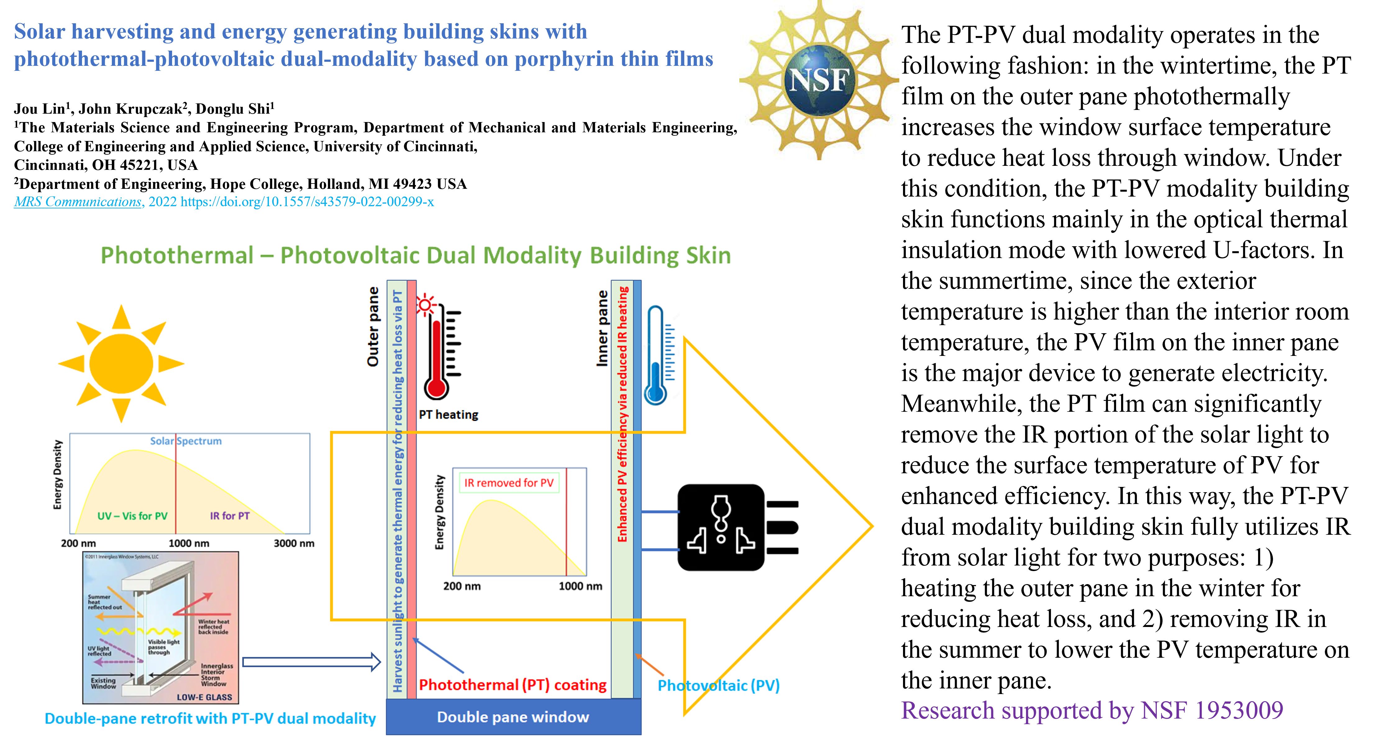
Solar harvesting and energy generating building skins with photothermal-photovoltaic modality based on porphyrin thin films
|
( MRS Communications 2022)
|
|
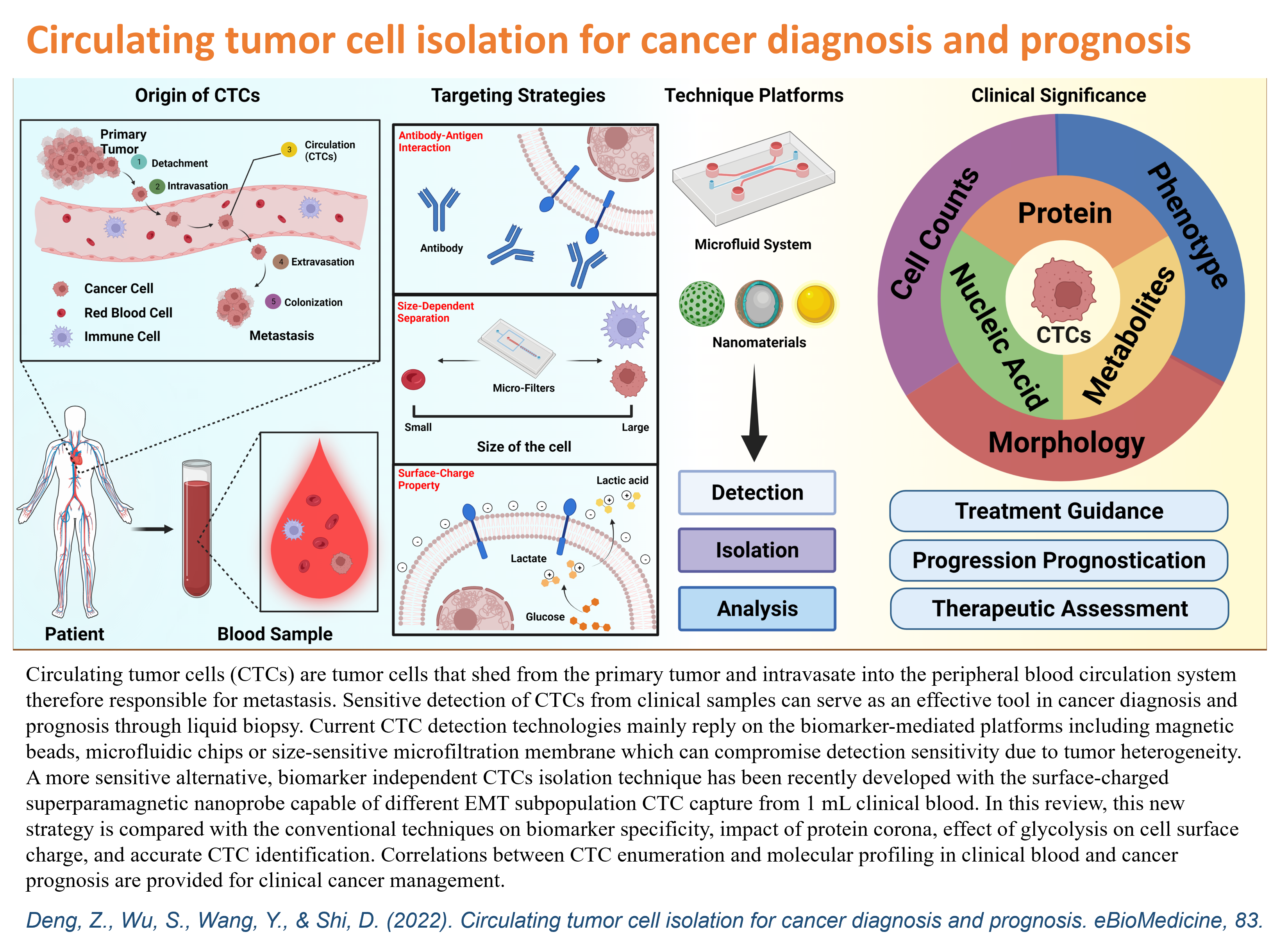
Circulating tumor cell isolation for cancer diagnosis and prognosis
|
(eBioMedicine 2022)
|
|
Transparent porphyrin-based hybrid films for spectral selective solar harvesting and energy generation
|
(Solar Energy Materials and Solar Cells 2022)
|
|
Entrapment of Airborne Particles via Simulated Highway Noise-Induced Piezoelectricity in PMMA and EPDM
|
(Energies 2022)
|
|
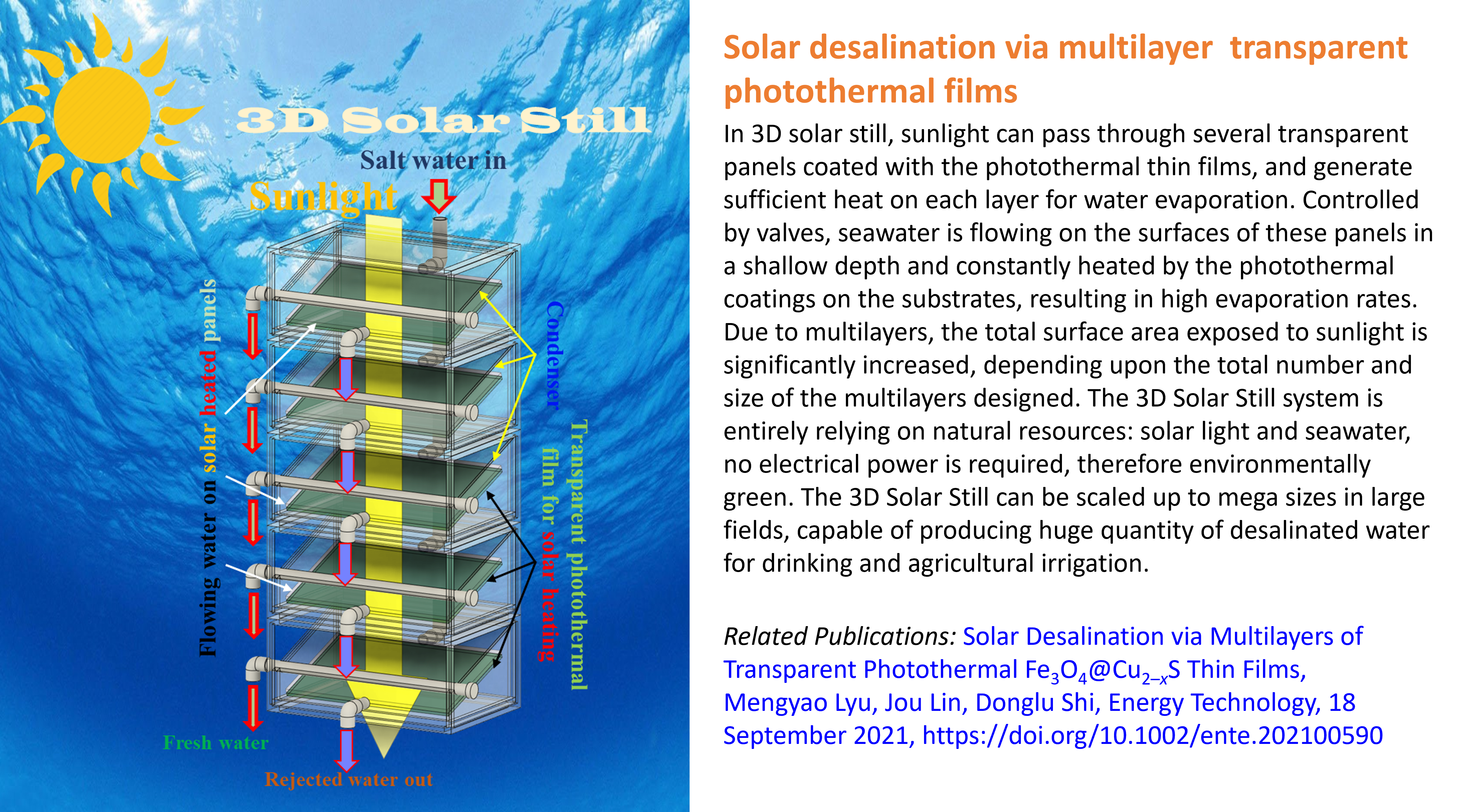
Solar Desalination via Multilayers of Transparent Photothermal Fe3O4@Cu2-xS Thin Films
|
(Energy Technology 2021)
|
|
Dual targeting with cell surface electrical charge and folic acid via superparamagnetic Fe3O4@Cu2-xS for photothermal cancer cell killing
|
(Cancers 2021)
|
|
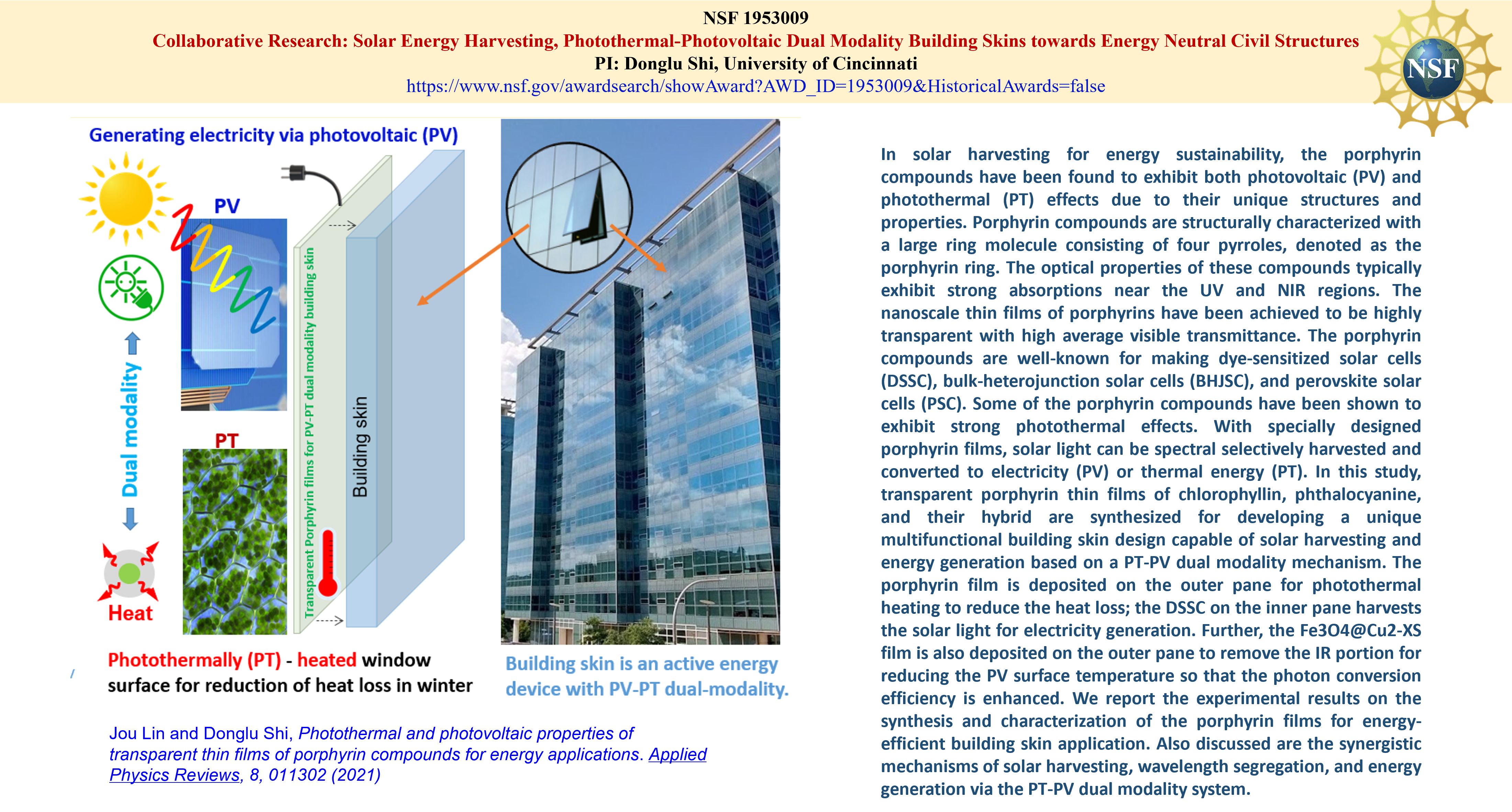
Photothermal and photovoltaic properties of transparent thin films of porphyrin compounds for energy applications
|
(Applied Physics Reviews 2021)
|
|
Solar harvesting through multilayer spectral selective iron oxide and porphyrin transparent thin films for photothermal energy generation
|
(Advanced Sustainable Systems 2021)
|
|
Nanoparticle Delivery Systems with Cell-Specific Targeting for Pulmonary Diseases
|
(American journal of respiratory cell and molecular biology 2021)
|
|
How effective is a mask in preventing COVID-19 infection?
|
(Medical devices & sensors 2021)
|
|
Photonically-Activated Molecular Excitations for Thermal Energy Conversion in Porphyrinic Compounds
|
(The Journal of Physical Chemistry C 2020)
|
|
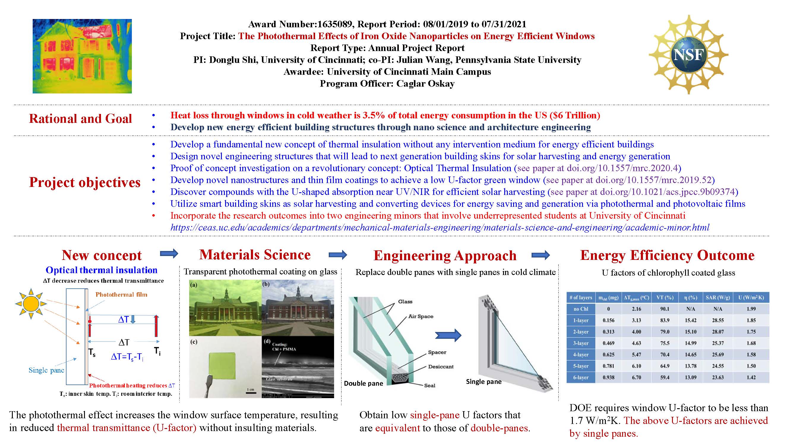
Optical thermal insulation via the photothermal effects of Fe3O4 and Fe3O4@Cu2-xS thin films for energy-efficient single-pane windows
|
(MRS Communications 2020)
|
|
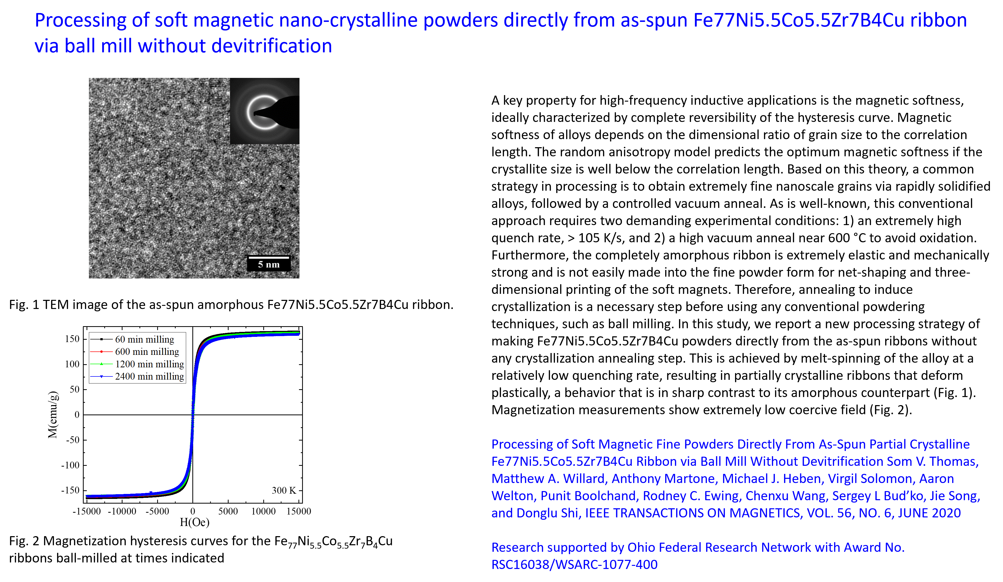
Processing of soft magnetic fine powders directly from as-spun partial crystalline Fe77Ni5.5Co5.5Zr7B4Cu ribbon via ball mill without devitrification.
|
(IEEE Transactions on Magnetics 2020)
|
|
Light angle dependence of photothermal properties in oxide and porphyrin thin ?lms for energy-ef?cient window applications.
|
(MRS Communications 2020)
|
|
Nanoparticle Delivery of Proangiogenic Transcription Factors into the Neonatal Circulation Inhibits Alveolar Simplification Caused by Hyperoxia.
|
(American journal of respiratory and critical care medicine. 2020)
|
|
The S52F FOXF1 Mutation Inhibits STAT3 Signaling and Causes Alveolar Capillary Dysplasia.
|
(American journal of respiratory and critical care medicine. 2019)
|
|
Effective reduction of building heat loss without insulation materials via the
photothermal effect of a chlorophyll thin film coated "Green Window"
|
(MRS Communications 2019)
|
|
Highly Efficient In Vivo Targeting of
the Pulmonary Endothelium Using Novel Modifications of Polyethylenimine: An Importance of Charge
|
(Advanced Healthcare Materials 2018)
|
|
"Minimalist" Nanovaccine Constituted from
Near Whole Antigen for Cancer Immunotherapy
|
(ACS Nano 2018)
|
|
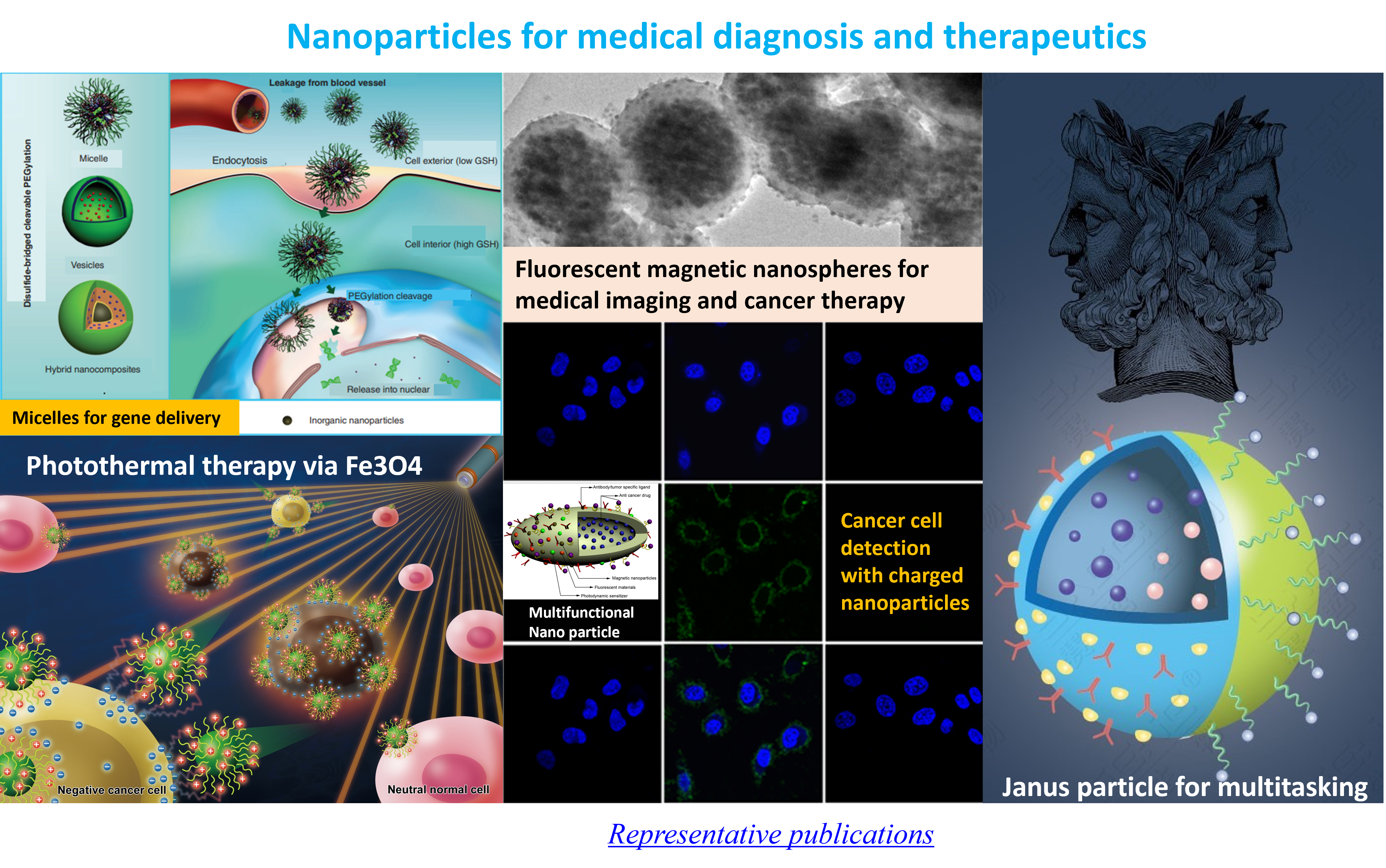
Nanomaterials for Cancer Precision Medicine
|
(Advanced Materials 2018)
|
|
Fever-Inspired Immunotherapy Based on Photothermal CpG
Nanotherapeutics: The Critical Role of Mild Heat in Regulating Tumor Microenvironment
|
(Advanced Science 2018)
|
|
Photothermal effect on Fe3O4 nanoparticles irradiated
by white-light for energy efficient window application
|
(Solar Eng. Mat. & Solar Cells 2017)
|
|
Biomarkerless targeting and
photothermal cancer cell killing by surface-electrically-charged
superparamagnetic Fe3O4 composite nanoparticles
|
(Nanoscale 2017)
|
|
Targeting and Regulating of an
Oncogene via Nanovector Delivery of MicroRNA using Patient-Derived Tumor
Xenografts
|
(Theranostics 2017)
|
|
Targeting
Negative Surface Charges of Cancer Cells by Multifunctional Nanoprobes
|
(Theranostics 2016)
|
|
A
Graphene Quantum Dot (GQD) Nanosystem with Redox-Triggered Cleavable PEG Shell Facilitating Selective Activation of Photosensitiser for Photodynamic Therapy
|
(RSC Advances 2016)
|
|
A
Multimodal System with Synergistic Effects of Magneto-Mechanical, Photothermal, Photodynamic and Chemo Therapies of Cancer in Graphene-Quantum Dot-Coated Hollow Magnetic Nanospheres
|
(Theranostics 2016)
|
|
Photo-fluorescent
and Magnetic Properties of Iron Oxide Nanoparticles for Biomedical Applications
|
(Nanoscale 2015)
|
|
Disulfide-Bridged
Cleavable PEGylation in Polymeric Nanomedicine for
Controlled Therapeutic Delivery
|
(Nanomedicine
2015)
|
|
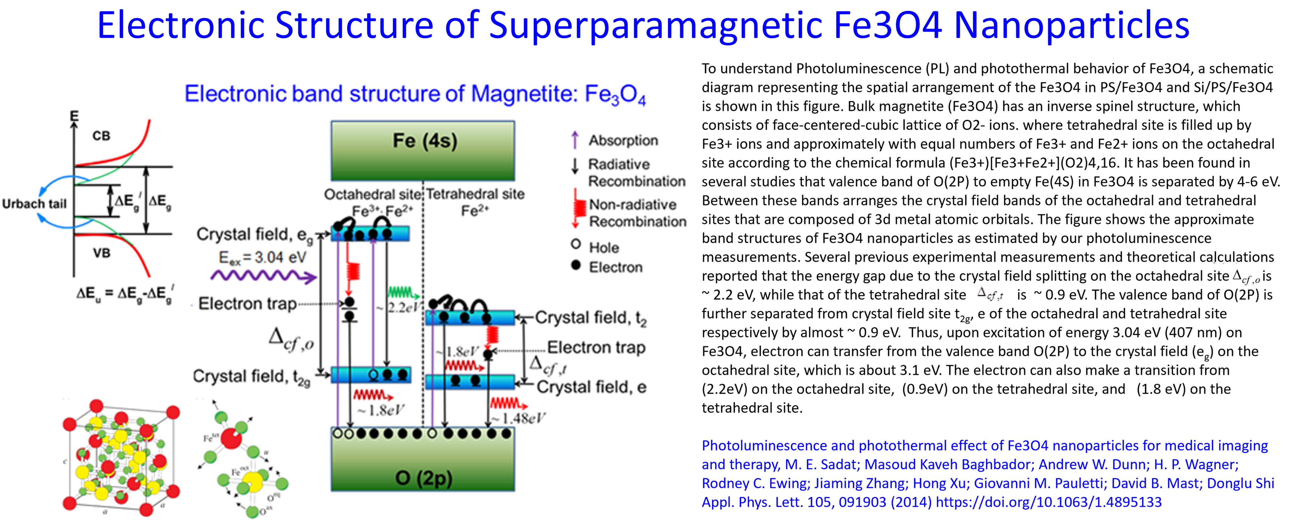
Photoluminescence
and Photothermal Effect of Fe3O4 Nanoparticles for Medical Imaging and Therapy
|
(App.
Phys. Lett. 2014)
|
|
Dual
Surface-Functionalized Janus Nanocomposities of
Polystyrene/Fe3O4@SiO2 for Simultaneous
Tumor Cell Targeting and Stimulus-Induced Drug Release
|
(Adv. Materials 2013)
|
|
A Versatile
Multicomponent Assembly via β-cyclodextrin Host-Guest Chemistry on Graphine
for Biomedical Applications
|
(Small
2012)
|
|
Engineered Redox-Responsive PEG Detachment Mechanism in PEGylated Nano-Graphine Oxide
for Intracellular Drug Delivery
|
(Small
2012)
|
|
Engineered
Multifunctional Nanocarriers for Cancer Diagnosis
and Therapeutics
|
(Small
2011)
|
|
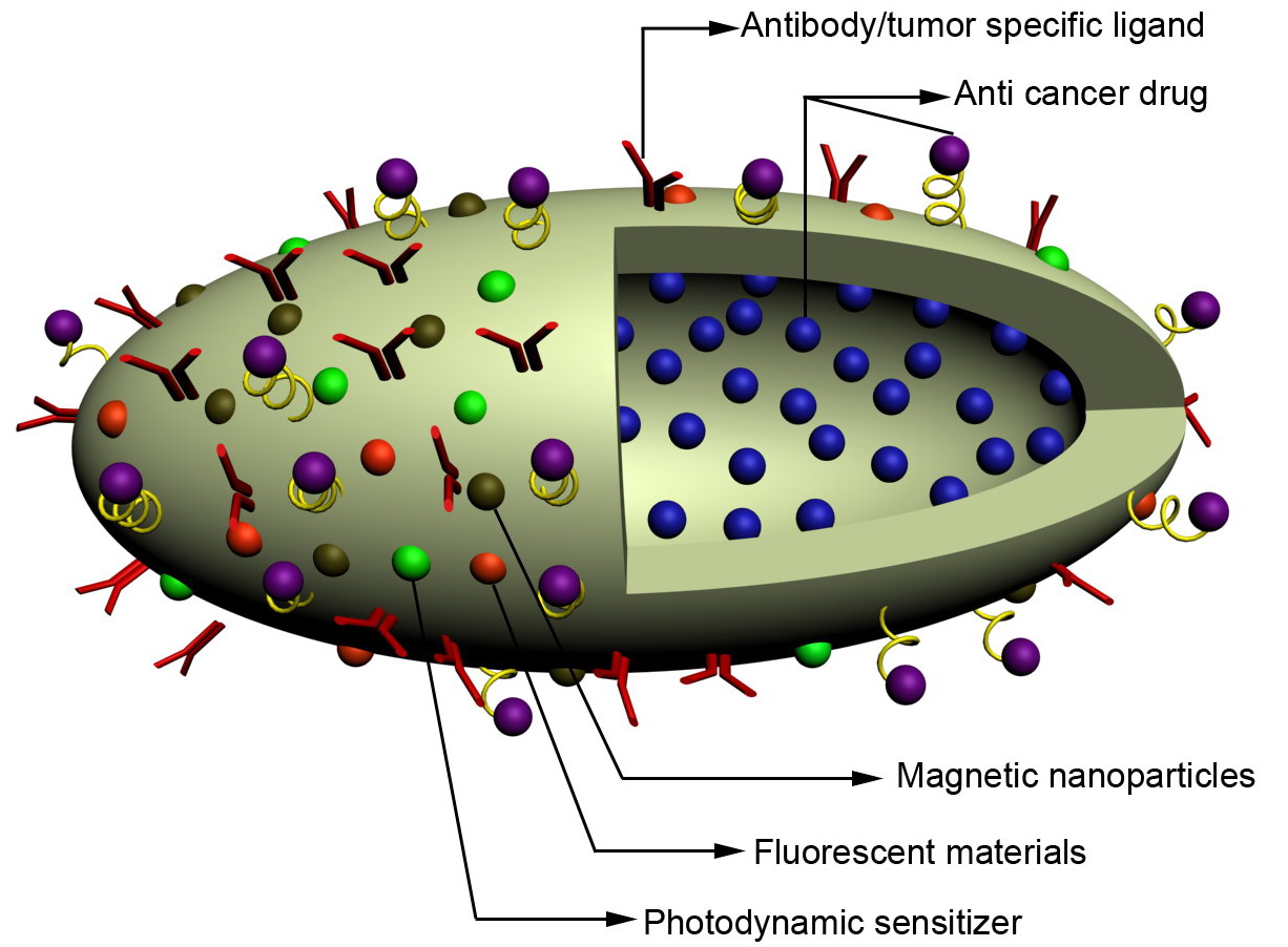
Fluorescent,
Superparamagnetic Nanospheres for Drug Storage, Targeting, and Imaging: A
Multifunctional Nanocarrier System for Cancer
Diagnosis and Treatment
|
(ACS
Nano. 2010)
|
|
Integrated
Multifunctional Nanosystems for Medical Diagnosis
and Treatment
|
(Adv.
Funct. Materials 2009)
|
|
Fluorescent
Polystyrene-Fe3O4 Composite Nanospheres for In Vivo Imaging and Hyperthermia
|
(Adv.
Materials 2009)
|
|
5f-6d
orbital hybridization of trivalent uranium in crystals of hexagonal symmetry: Effects on electronic energy levels and transition intensities
|
(Phys. Rev. B 2009)
|
|
In vivo Imaging and Drug Storage
by Quantum-Dot-Conjugated Carbon Nanotubes
|
(Adv.
Funct.
Materials 2008)
|
|
Nanoscale Solute Partitioning in Bulk Metallic Glasses
|
(Adv.
Materials 2008)
|
|
Effects
of plasma surface modification on interfacial behaviors and mechanial properties of carbon nanotube-Al2O3 nanocomposites
|
(Appl.
Phys. Lett. 2007)
|
|
Neutron diffraction study of the structure and low-temperature
phase transformation in ternaty NiAl + M (M=Ni, Fe, Co) allows
|
(Scripta Materialia 2007)
|
|
In Vivo Imaging by Luminescent Nanotubes
|
(Adv.
Materials 2007)
|
|
Luminescent
Carbon Nanotubes by Surface Functionalization
|
(Adv.
Materials 2006)
|
|
Processing
Dependence of Texture, and Critical Properties of YBa2Cu3O7-δ Films on RABiTs Substrates by a Non-Fluorine MOD Method
|
(J. Am. Ceram. Soc. 2006)
|
|
Strontium-Induced
Oxygen Defect Structure and Hole Doping in La2-xSrxCuO4
|
(Phys. Rev. Lett. 1991)
|
|
Synthesis,
structure and superconductivity in the Ba1-xKxBiO3-y system
|
(Nature 1988)
|